
Deep or eloquent-area lesion
A lesion near areas controlling speech, movement, vision or other critical functions may be too risky for immediate open removal.
A stereotactic biopsy uses MRI or CT-based 3D navigation to reach a deep or difficult brain lesion and collect a small tissue sample. The pathology result helps your doctors identify the disease and choose the right treatment — without requiring a large craniotomy in many cases.
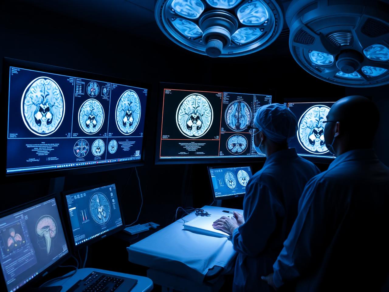
A stereotactic brain biopsy is a minimally invasive operation in which a neurosurgeon uses a frame-based or frameless navigation system to calculate a three-dimensional route to a brain lesion. Through a small burr hole, a narrow biopsy needle removes several tiny samples. A neuropathologist then studies the cells, and may perform immunohistochemistry, molecular profiling or infection tests.
It is primarily a diagnostic procedure, not a treatment that removes the whole lesion. The result can distinguish a tumour from lymphoma, infection, inflammation, demyelination or another condition and can materially change the treatment plan.
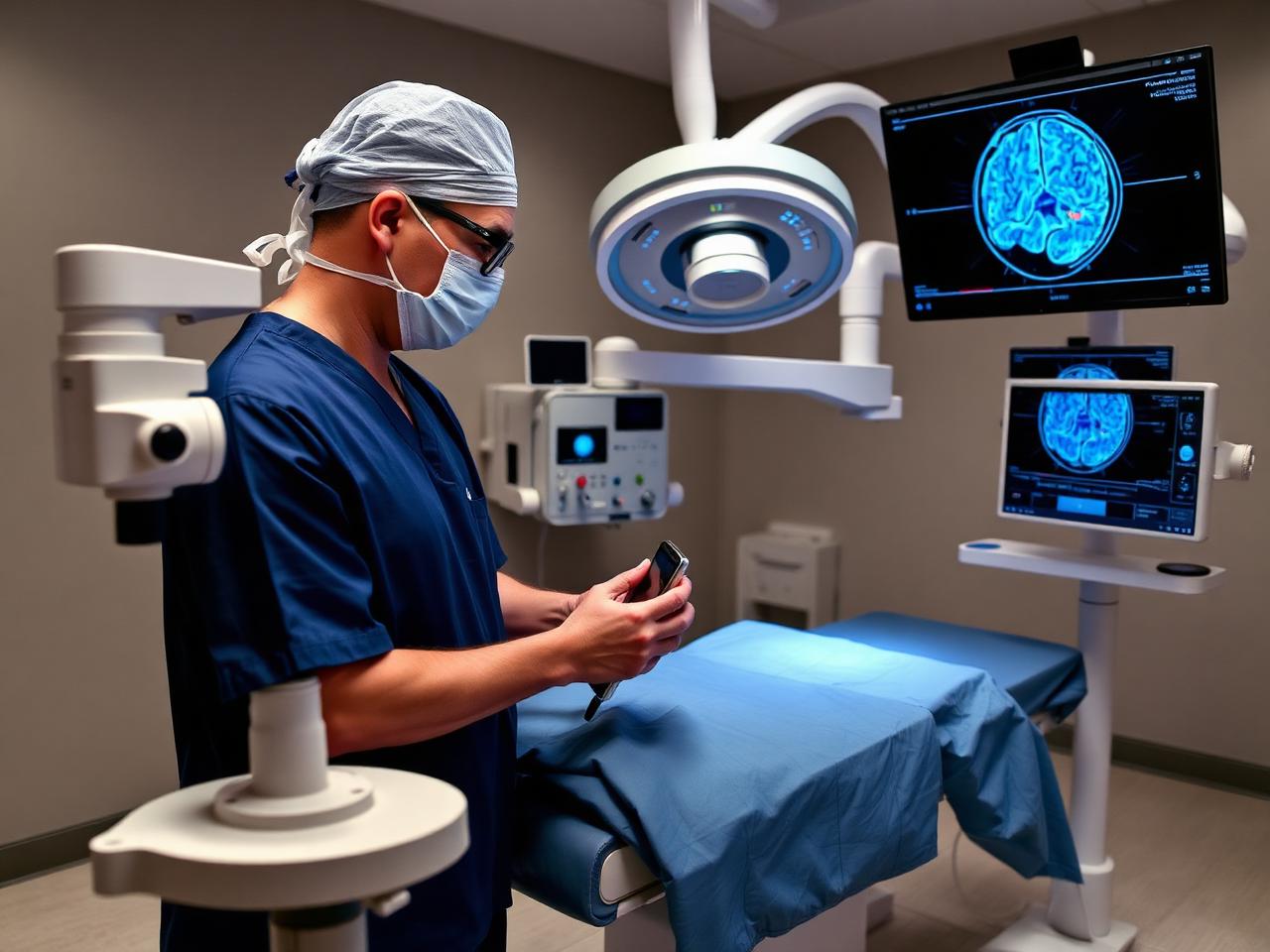
The surgeon combines current MRI/CT imaging with navigation planning before selecting the safest trajectory.
The decision is made after reviewing your symptoms, MRI/CT findings, medical history and whether a safer or more useful diagnostic route exists.
A lesion near areas controlling speech, movement, vision or other critical functions may be too risky for immediate open removal.
Imaging cannot reliably distinguish glioma, lymphoma, metastasis, infection, inflammation or demyelinating disease.
A representative sample may identify the disease and avoid several larger operations.
When complete removal is not feasible, tissue can establish type, grade and molecular markers for targeted planning.
Biopsy is often essential because treatment is usually nonsurgical; steroids can obscure pathology and must be discussed urgently.
Age, health conditions or lesion location may make a minimally invasive diagnostic approach preferable.
Neurosurgical examination; MRI with contrast, blood tests and medication review. Blood thinners may require a supervised pause.
A stereotactic MRI/CT dataset maps the target, blood vessels and safest route. Frame-based or frameless guidance is selected.
Under general anaesthesia or sedation, the surgeon makes a small incision and burr hole, then advances the needle to the target.
Several samples are obtained. Frozen-section review may confirm that diagnostic tissue is present before closure.
Neurological checks and often a CT scan look for bleeding. Pathology guides the next multidisciplinary treatment discussion.
Best when a tissue diagnosis is needed and removing the lesion is unsafe, unnecessary or unlikely to help immediately.
May be chosen when more tissue is required, pressure must be relieved, or safe removal can provide both diagnosis and treatment.
Advanced MRI, PET, spectroscopy, blood/CSF testing or interval imaging may be appropriate when biopsy risk exceeds likely benefit.
Prices are planning ranges, not guarantees. Currency conversion and final hospital billing can vary.
| Care component | Indicative range | Common inclusions |
|---|---|---|
| Standard stereotactic biopsy package | USD 3,500–7,000 approx. ₹2.9–5.8 lakh | Surgeon, operating room, anaesthesia, navigation and standard stay |
| Advanced pathology / molecular tests | Case-dependent | IHC, tumour markers, microbiology or genomic profiling |
| Extended ICU or hospital care | Case-dependent | Needed only when clinical condition or complications require it |
Get an expert second opinion or learn about medical travel support in India. You may also explore our specialist network, brain tumour surgery and radiation therapy.
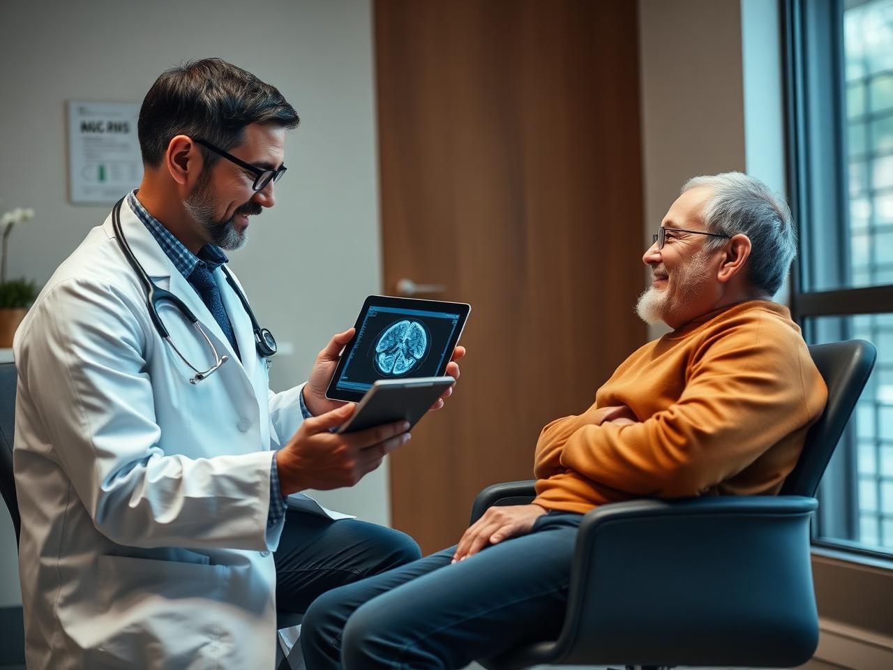
Expect neurological observation, wound checks and mild scalp discomfort. A post-procedure scan may be performed. Do not drive until your doctor clears you.
Many patients gradually resume light activities. Keep the wound clean, take medicines exactly as prescribed and attend the pathology review.
New weakness, speech or vision change, worsening severe headache, repeated vomiting, seizure, confusion, fainting, fever or wound leakage needs immediate assessment.
A rigid stereotactic frame supplies coordinates with high mechanical accuracy.
Optical or electromagnetic tracking guides instruments without a traditional frame.
A robotic arm may align the planned trajectory and support instrument placement.
CT planning or intraoperative CT can assist selected targets.
MRI supplies detailed soft-tissue definition for many lesions.
For selected ventricular lesions, endoscopy may obtain tissue and restore CSF flow.
These are possibilities investigated — not conditions that MRI alone confirms.
Tissue defines histology, grade and relevant molecular markers.
Often needs tissue before treatment; steroid timing matters.
Biopsy may identify tumour type when no primary cancer is known.
Samples can be tested for bacterial, fungal, parasitic or tuberculosis infection.
Selected cases of vasculitis or neurosarcoidosis may require tissue.
Tumefactive demyelination can closely mimic a tumour.
The result should guide therapy, surveillance or surgery.
The target must be reachable while reducing risk to vessels and critical pathways.
If safe removal can also treat the disease, open surgery may offer more benefit.
Platelets, clotting, blood pressure and blood-thinning medicines are reviewed.
Heart, lung and general health guide the anaesthesia plan.
The patient understands alternatives, limitations and neurological risks.
Matched by lesion location, navigation experience and reasonable alternatives.
DICOM images support decisions about trajectory and whether biopsy is appropriate.
Frame-based, frameless and suitable image-guided or robotic options.
Neuropathology, immunohistochemistry, microbiology and molecular markers.
Hospital-specific planning ranges with likely exclusions stated.
Appointments, admission, travel, language, discharge and follow-up.
Send DICOM MRI, reports, symptoms and current medicines on WhatsApp.
Neurosurgeon reviews imaging, hospital options and a written estimate.
Visa invitation letter, appointments, accommodation and airport pickup.
Pre-operative tests, the procedure, neurological monitoring and discharge.
Report review, multidisciplinary treatment plan and follow-up coordination.
Support across complex treatment journeys in India.
Patients from India and more than 20 countries.
Hospitals with neurosurgery, neuro-ICU, imaging and pathology.
Reports organised and plans communicated clearly.
Interpreters and local assistance according to need.
Discharge and pathology records coordinated for review.
Selection depends on surgeon, technology, pathology, ICU capability, budget and availability.
Tertiary neurosurgery, imaging, critical care and oncology.
Integrated neuroscience and cancer-care services.
Neurosurgery, critical care and imaging.
Multi-city neurosciences and multidisciplinary care.
Neurology, neurosurgery, radiology and critical care.
Neuroscience, oncology, imaging and rehabilitation.
No. Infection, inflammation, demyelination, lymphoma, primary tumours and metastases may resemble one another. Specialist review comes before biopsy.
Not always. When diagnosis is the immediate goal and the lesion is deep or near critical areas, stereotactic biopsy may obtain tissue through a small burr hole.
MRI or CT is loaded into navigation software to plan the target and a route avoiding vessels, ventricles and functional structures.
Specialist pathology improves diagnostic yield, but repeat biopsy or open surgery is occasionally needed.
Medifly coordinates hospital options, visa documents, accommodation, transfers, interpreters and follow-up.
A written estimate explains expected charges and potential extras such as advanced molecular tests.
Educational examples — not testimonials or guaranteed outcomes.
Biopsy may be considered when removal carries high functional risk.
Imaging, steroid exposure and tissue timing are coordinated.
A representative lesion may establish tumour type.
Tissue is divided for pathology and microbiology.
Molecular information guides radiation and drug treatment.
Biopsy proceeds only if tissue is likely to change management.
Compare biopsy, resection and observation before deciding.
Your centre lacks suitable stereotactic technology.
The case requires neuropathology or molecular profiling.
Your doctor advises that diagnosis should not be delayed.
Compare an itemised Indian estimate including travel.
One team coordinates visa, admission and follow-up.
An indicative package is USD 3,500–7,000 (approximately ₹2.9–5.8 lakh), depending on hospital, navigation technology, lesion location, anaesthesia, ICU needs and pathology tests. A personalised written estimate is needed.
It may be performed under general anaesthesia or under local anaesthesia with sedation. The choice depends on the technique, target, patient condition and the neurosurgical team's protocol.
Diagnostic yield is generally high at experienced centres, but no biopsy is guaranteed. Small, necrotic, heterogeneous or difficult lesions can produce insufficient or nonrepresentative tissue.
Many patients stay one to three nights and resume light activity within about one to two weeks, although recovery varies with lesion location, symptoms and complications.
It is a minimally invasive neurosurgical procedure performed through a small scalp incision and burr hole, but it still carries meaningful risks such as bleeding, seizure, infection and neurological deficit.
A preliminary result may be available in a few days. Final diagnosis can take longer when immunohistochemistry, molecular profiling, cultures or outside review is needed.
Tumour seeding along a stereotactic biopsy tract is considered very rare. The more immediate concerns are bleeding, seizure, infection and neurological injury, which your surgeon should quantify for your case.
No ethical medical coordinator can guarantee either. Medifly can arrange specialist review, a written hospital estimate and end-to-end logistics; clinical decisions and outcomes depend on the treating team and the patient's condition.
Often around 7–14 days is practical for consultation, pre-operative testing, biopsy, observation and preliminary pathology, but the neurosurgeon may advise longer based on symptoms and results.
Medifly will coordinate review by an experienced neurosurgeon, suitable hospital options, an indicative treatment plan and a written estimate. For emergencies or rapidly worsening neurological symptoms, contact local emergency services immediately.